Thursday, December 10, 2015

Thanks to the Deathstar

This week was pretty hectic working on our SSTEM projects as well as our final draft. I tried running some more gels this week in hopes of seeing some DNA from my newest extractions. My gel still did not show any DNA from under the UV lighting. In my presentation I felt a bit afraid of showing gels without polymorphic banding, but my mentors/SSTEM scholars reassured me that it was not a waste. Through my failure of not extracting DNA from the cholla spines actually proved those protocols I used were not effective and to avoid using the same steps next time.

 One of my gels from this past week that showed I had not yielded any DNA from my extractions. In my next post I hope to have some polymorphic banding.

Thursday, December 3, 2015

Going Forward On A Speeder Bike


Fortunately, I was able to conduct these DNA extractions in the biology lab and I was able to retrieve a piece of cylindropuntia bigelovii from the PC greenhouse. The next steps will be to create different reagents in order to extract the DNA from the cholla spines. I also plan on collaborating with other DNA plant extractions I have read from previouse S-STEM scholars in hopes of having some luck on extracting DNA and seeing it under the UV light after electrophoresis. I need to see polymorphic banding from cholla cacti in order to answer my question on whether chollas reproduce sexually or asexually.

Thursday, November 19, 2015

Stuck in the Deserts of Tatooine

During the week of November 19th, I was still working on my extractions and began to run gels on them. I figured that if I had my samples incubate overnight in the lysis buffer the fatty membranes and proteins associated with the DNA would have broken down. I also thought I would have seen a DNA pellet form in the -20 degree isopropyl-alcohol after waiting 30 minutes, but I was not able to see anything form. After extracting most of the isopropyl-alcohol from the microcentrifuge-tube, I allowed it to air-dry underneath the hood. I then suspended the DNA in distilled water. My samples underwent electrophoresis and here is the gel:
 This gel held three samples from my three separate DNA extractions. I was not able to see any DNA under the UV light. It was not something I had hoped to see.

Thursday, November 12, 2015

Beginning Life on Alderaan


During the week of November 12, I continued trying to perfect my Cacti DNA extraction. This new protocol involved my older protocol to extract DNA from ants. This protocol involved:
  • I put 5-15 spines into a microtube and wash with bleach.
  • I then decant and rinse the spines twice with distilled water.
  • I cut the spines into smaller pieces with nail-cutter scissors.
  • I add 90 micro-liters of nuclease free water, 1 micro-liter of 10% SDS, 2 micro-liters of 20% meat tenderizer solution.
  • I heated the tube at 75 degrees Celsius for 10 minutes in a water bath and agitated the tube thereafter with a flick of my finger.
  • I heated the tube at 95 degree Celsius for 5 minutes in a heating block.
  • I agitate the tube with a flick of my finger then allow the tube to incubate in room temperature overnight.
  • I take the sample and put it into the centrifuge for 13.2 rpm for 5 min.
  • I take the sample out of the centrifuge and remove the supernatant from the top in order to transfer into a new microcentrifuge-tube. 
  • I add -20 degrees Celsius isopropyl-alcohol equal to the amount of supernatant and put the tube at -20 degrees Celsius for 30 minutes.
  • I centrifuge the sample and collect the supernatant into a new tube.
The heating block used and the other picture shows two samples with -20 degree Celsius isopropyl-alcohol.

Thursday, November 5, 2015

Beginning A Trek Into A New Naboo

During the week of November 1st, I began the protocol for extracting DNA from the cholla cacti. I received a piece of cholla from our green house in hopes of extracting DNA and finding clear polymorphic banding after running the DNA sample in a gel. There were various ways on how I began isolating the DNA.

  • The first protocol I used consisted of washing 5-15 spines in a microtube and washing them in bleach and decant with a disposable pipet.
  • I rinse the spines with distilled water twice and decant.
  • I cut the spines with nail-cutter scissors.
  • I added 1000 micro-liters of lysis buffer after the spines are cut and incubate for 30 minutes at 65 degrees Celsius. 
  • I disrupt the spines by vortex machine and incubate for another 30 minutes.
  • I then incubate the sample overnight by allowing it to sit at room temperature.
  • I add 5 drops of protease to the microcentrifuge-tube and heat for 10 minutes at 65 degrees Celsius.
  • I vortex the sample and put it in the centrifuge at 13.2 rpm for 4.0 minutes.
  • I take the sample out of the centrifuge and remove the supernatant from the top in order to transfer into a new microcentrifuge-tube. 
  • I add -20 degrees Celsius isopropyl-alcohol equal to the amount of supernatant and put the tube at -20 degrees Celsius for 30 minutes.
  • I centrifuge the sample and collect the supernatant into a new tube.

Thursday, October 29, 2015

Two Suns for Tatooine


During the week of October 25th, I worked on figuring out what kind of protocol to use in order to do the cactus spine DNA extraction and I was also working on finding sources for the final paper. Matt happened to lend me some books such as A Natural History of the Sonoran Desert, Environmental Biology of Agaves and Cacti, and Cacti Biology and Uses. The paper will be about how I was able to extract DNA from a cholla and how I was able to distinguish the polymorphic banding. I hope the protocol I use will be able to produce a high yield of cholla DNA. 


Thursday, October 22, 2015

A New Republic

This week I began with running a new PCR with primer 4 in order to get clear banding and making fresh gels for electrophoresis. I was able to run four gels yesterday and I looked at the results. Unfortunately, they did not come out the way I had expected. The ladder did not come out as clear as the others had and the bands were just as sloppy. I will be running new gels today in order to see if it was the total amount of time running the gels that had affected my results. I will most likely change the time of how long I want my gels to run.

Colony A Primer 4 gel

 Colony B Primer 4 gel
Colony C Primer 4 gel 

Colony D Primer 4 gel

Thursday, October 15, 2015

Figuring Out the Confederacy and Moving Further

I was looking at photos of my gels from the previous semester and I figured out why my bands were not coming out as great as they should. The best primer to use for amplification of my ant DNA should be primer 4. The photos of the bands amplified from primer 4 are the most clear.
Here is the photo compared to the primer I was using.

The primer I was using.

 
The primer I will be using from now on.


This week I was mainly working on my research proposal that I did not get to run the new PCR, but I will as soon as I can. My proposal dealt with my new project on extracting DNA from
Cylindropuntia bigelovii (teddy bear cholla). I cannot wait to start the cacti DNA extraction protocol. I'll be super busy in the lab even more from now on!


Thursday, October 8, 2015

Reaching Endor

I am still analyzing my gels in order to see what is the problem with not getting clear banding, but I have also started my research on extracting DNA from plants. I will begin extracting DNA from cholla in hopes of seeing similar banding in each sample of cholla. I also began planting bonsai trees. I planted a Japanese Red Maple and a Katsura. If they germinate soon, I might just try to extract DNA from those little trees as well! Below are the pictures of the bonsais I have recently planted in the lab:

Japanese Red Maple Bonsai


Katsura Bonsai (Has a cotton candy smell.)


Thursday, October 1, 2015

Solving The Mystery of the Running Speeder Bikes

This week I ran another PCR because Matt thought it would be better than nothing. I was not able to understand why I was not receiving any bands in my gels. I made some fresh gels yesterday and ran my fresh amplified DNA in hopes to see if there would be some polymorphic bands. To my astonishment/relief, there were bands! But now there is a new mystery to solve. The gels seemed to have stars in them when put under the UV light. We are not sure if this was due to the DNA running off in little fragments or maybe the agarose was not cooked fully. We are all on the case and I will try my best to figure this out soon enough!


Tuesday, September 29, 2015

DNA on Speeder Bikes or so I Thought

This week I am working on making more 2% Agarose gels in order to run some with my newly amplified Ant DNA. I ran the PCR last Thursday, and I am hoping for the bands in the gels to be clear. I need the bands to be clear in order to make a concise measurement of whether certain bands are similar to one another.
I ran my gel with the amplified DNA and there were no bands! I then ran another gel with my regular DNA samples and saw polymorphic banding. I wanted to make sure I actually had DNA in my samples and wanted to know why there was no DNA showing in the gels from the PCR. I will be running another gel next week on the same amplified DNA samples. I need to figure this one out.


Wednesday, September 16, 2015

The Old Republic

This week I began making new gels with 2% Agarose in order to see if the polymorphic bandings from the ants would seem clearer than the bands in a gel of 1% Agarose. It did not seem to work and I am wondering if I should just run another PCR on my ant DNA. I ran three gels with 6 microliters of DNA and 2 microliters of the orange loading dye. It occurred to me, after I reviewed my previous gels over the summer, there was barely enough DNA shown in the first gel from my amplified DNA samples. Here are the first three gels I had ran a couple of weeks ago from the first PCR in a 1% Agarose Gel:

Colony A

 
 Colony B
 
 Colony C
 
In the Colony A gel it shows barely any DNA, and when I ran a gel yesterday with the same DNA, but in a 2% Agarose gel, nothing showed up. I felt as though it was a waste of three 2% Agarose gels.

My poor agarose gels.

Monday, September 14, 2015

Storm Troopers on the Rise


I am happy to be back this Fall 2015 Semester! Good wishes to you all on this new semester! I'd like to share with you all the most interesting thing I have been researching so far about ants. I've been researching their many different adaptations. One of my favorite adaptations of an ant species is how they are capable of stealing another specie’s colony in order to survive. They kill the colony’s queen and all of her children. The tend to leave some of the larva alive in order to keep them as their slaves. This ant species is known as Polyergus breviceps or Slavemaker ants. They are basically ruthless serial killers.
Here’s a video of the ants about to embark into new territory:
 

Tuesday, May 5, 2015

The Never-ending Droids

In the week of April 30th I was finished with my DNA extractions and began running a PCR. I used four different primers. I had five DNA extractions in total due to the five different pharaoh ant colonies. I ran all five samples separately for each primer. After the PCR, I began running a gel from each primer set. I found a trend in the polymorphic bands while looking at the gel under the UV lighting. It seemed as though colonies 1&3 had similar polymorphic bands while colonies 2,4,&5 had similar polymorphic banding with one another. Below you shall see the gels of each different primer and the DNA amplification of each colony over five lines.

Gel with DNA samples amplified with Primer 1.


Gel with DNA samples amplified with Primer 2.


Gel with DNA samples amplified with Primer 3.


Gel with DNA samples amplified with Primer 4.


Monday, May 4, 2015

Running Trade Federation Gels

In the week of April 16th I ran gels on the DNA samples I amplified with the PCR. It did not turn out very well;I was a bit disappointed in how the amplification turned out. I wanted to see bright,beautiful polymorphic bands, but only recieved unclear, smeared remnants of my DNA in the gel. I had used primers 1 & 2 to amplify two sets of samples. I used four samples of DNA with primer 1; one control DNA extraction and four other DNA extractions with a novel method. I did the same samples for primer 2 as well. When I ran a gel on each primer to see which primer would amplify the DNA better, I did not see good results. Below are the pictures of the gels and how they turned out. 


Gel of amplified DNA with primer 1.

Gel of amplified DNA with  primer 2.

Monday, April 27, 2015

Amplification of the Trade Federation

The week of April 9th was one filled with new techniques. Matt helped me learn how to run a PCR! I was excited to see if the DNA samples I had would be amplified . The process was not as hard as actually doing a DNA extraction. After I was done running the PCR, I was taught how to run a gel with my amplified DNA which is different from the usual electrophoresis with non-amplified DNA. Aftter I ran my gel and looked at it under the UV lighting it seemed as though the primers had done well. I used both primers instead of just one each for my DNA samples. Below is a picture of how well both primers worked with my DNA extractions.

This is a gel containing both primers 1&2 from my first PCR.

Thursday, April 23, 2015

On the Planet Genesis

In the week of April 23rd I was working on collecting ants from five locations on PC campus. I was happy to find five pharaoh ant colonies. I am pretty sure I looked like a crazy person scooping up ants into little eppendorf tubes with tweezers. I needed enough ants to have 25 extractions. I was planning on having five control extractions with E.Z.N.A. Insect DNA kit protocol and 20 others with the novel method. I happened to finish all the extractions in two days and ran a gel to make sure I had DNA. Below is a picture of the gel I ran with a couple of DNA samples.

The control sample from the E.Z.N.A. Insect DNA protocol.

The DNA samples from the novel method.

Not much DNA from the pharaoh ants, but there was enough to run a PCR.

Thursday, April 2, 2015

The Fake Royal Naboo

I  started with three new DNA extractions with the E.Z.N.A Insect kit protocol and I ran a gel with the three new DNA samples. I put the gel under the UV lighting and there was actually some DNA! There was not as much DNA as the first protocol I used, but the E.Z.N.A protocol delivered enough DNA that there could be a chance of doing a PCR with it. I am excited to move forward with these results and see where it will take me.
                                    
 
Earlier this week was an adventure out to the northside of PC's campus! We were going to dig up an ant hill and find the queen. We ended up digging into plenty of dirt with the shovel and eventually piling it into the bucket. It was undetermined if we had secured the queen or not. When we got back in the lab we put the contents of the bucket into a TLC chamber. It is unclear if we have the queen at the moment, but it seems as though we do since the ants are now creating mini chambers for the larvae. It's pretty amazing how they create so many tunnels.

The established home for the ants.


Looking at larvae under a dissecting scope.


Tuesday, March 24, 2015

Down with the Trade Federation

I have begun the E.Z.N.A Insect DNA Kit Protocol to see if it matches up to the previous protocol conducted in the last DNA extraction. I will see if this protocol will yield more DNA and this protocol will be a control as well. I did electrophoresis on the new DNA and we tried looking at it under a UV lighting device, but it did not turn out the way we expected. I will have to run another electrophoresis in order to see the DNA bands again. It's still a mystery, but a mystery that can easily be solved!
                                              

On another note, Josh allowed me to use a dremel in order to cut plexiglass to make my ant farm! It was my first time working with a hand tool and I did not think I would be trusted with such a machine. I ended up cutting the glass and glued it together with a hot glue gun. Now all that needs to be done is putting some silicon on it.  

                                    
I can't wait to have my family of ants in there!

Monday, March 9, 2015

"Beam me up, Scotty!"

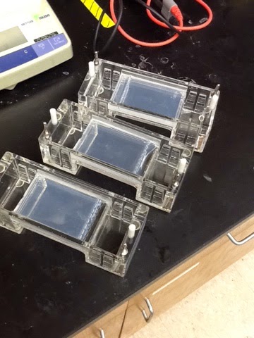
I have started on the new protocol this week! The one that actually harbors DNA at the end. I am positive the reason why the DNA was not seen under the UV lighting was due to a no heating process in the first protocol, so the DNA was not absolutely dissolved. Somehow the DNA beamed up out of the gel under the UV lighting with this new protocol containing the two heating processes. Josh danced for joy while Matt was impressed with the amount of DNA shown. I could not believe that I had collected so much DNA out of my extractions. All my thanks to such a wonderful protocol. 


I am now waiting to receive my primers to attempt on amplifying the ant DNA. I will be working on figuring out what specie of ant I have been collecting. I looked at my little furry friends under the dissecting-scope after defrosting them from the freezer. It turns out they have hair all over their bodies, otherwise known as setae, which are attached to nerves under their hard exoskeleton. They are to help them sense what is around them in their environment. I was also able to see their tarsi for which are the last several segments of the end of their legs. The claws at the tips of their legs are sharp enough to hold onto our skin, but too small to cause any harm.


I cannot wait to continue my research on the gene flow of ants. I think I will be also conducting DNA extractions from other insects as well. If you guys have any ideas of which insect species I should extract DNA from next, don't be afraid to mention it to me.


Here is another thing we happened to do in lab today with my fellow STEM Scholars Melisa, Bethany, & Zaira. In the STEM office, we had a red betta fish known as Olivia and she died due to a digestive block in her system. Matt assumed it was bacteria leading the way to the cause of her bloating. Bethany and I were too afraid to dissect her, so Melisa decided to dissect Olivia. Melisa (Dr. Frankenstein) had a blast while I held my nose looking through the dissecting microscope to see Olivia's guts. We were able to see the heart as well as leftover pellets (her food).

                                
                                    This is Olivia's heart.


Sunday, March 8, 2015

Attack of the Clones

I actually began my first electrophoresis! I was helped by wonderful Paul throughout the process. I learned how to put my 1 X TAE buffer together then my 1 X TAE buffer with 0.9 agarose. I had no idea agarose was the key ingredient to forming the gel in the gel rig. I thought it seemed pretty awesome. I had only one DNA sample for which I placed 2 microliters in the gel wells. When I was done running my gel I put it under the UV light. I also learned that the dye within my gel was light sensitive and could break down quickly resulting in not allowing me to view any DNA. It was a good thing I had a cardboard box. Unfortunately, we did not see any DNA, but this is only the beginning. I am sure I will be able to see some DNA in my next run of electrophoresis.
 

Here is my 1 X TAE buffer without the agarose of course.

Here is my gel undergoing electrophoresis under the cardboard box.
My gel with no result of DNA.

Thursday, March 5, 2015

Revenge of the Sith

I find that a DNA extraction does not work through some tedious protocol, but through magic! Just kidding! Thhe protocol I was using previously these past two weeks turned out to not serve me justice when looking under the UV lighting with my gels. Josh, my mentor, came up with a new protocol that turned out to result in actually exposing the DNA under the UV lighting after electrophoresis. The first protocol I used had no heating whatsoever, but in the new protocol there were two heating stages. I did however create a new DNA sample with the first protocol then used the new protocol. As before, after electrophoresis there was no DNA to be seen from the first protocol DNA sample. Below are pictures of two DNA samples from the new protocol under UV lighting after electrophoresis. 

Making some new gels under a cardboard box. ;)
I am about to prepare these gels with my DNA samples for electrophoresis.

I have just taken out the gel to view under UV lighting.

The first DNA sample I made with the new protocol under UV lighting.
Mine and Josh's DNA samples from the new protocol under UV lighting.
His DNA is shown from the second to last well in the gel.